
Can electrosmog exposure actually be detected? What does the blood picture look like and what does this mean for our health? Dark-field microscopy provides information. This variant of light microscopy for blood analysis , known for over 250 years. When this imaging is done, the blood cells appear bright on a dark background in greater detail, which has given it the name “dark field”. To analyse the blood to determine the state of health of a patient, a single drop of blood is taken. In some ways, it can be used as an early stage to help with a diagnostic of what might be present
This blog post talks about:
- what really the dark-field microscopy is and what it is used for;
- how can this method be beneficial for determining the level of electrosmog load;
- how it can help you recognise that the EMF protection measures you take are effective
What is dark-field microscopy?
Dark field microscopy and Enderlein therapy
The dark field microscopy method was developed in the 1920s by zoologist and bacteriologist Prof. Dr. Günther Enderlein (1872−1968). This is why the term Enderlein therapy is often used in connection with dark field microscopy.
The darkfield microscope is based on the theory of pleomorphism . There are microorganisms in the blood, such as cells, bacteria, viruses or fungi that develop depending on the condition of their environment. Any kind of disease requires a specific biological terrain, which dictates whether and what disease can occur. More highly developed microorganisms can only occur in a correspondingly poor environment and are therefore the consequence, and not the cause of a disease. According to Enderlein, these can be treated with different forms of these microorganisms.
On what principle is dark field microscopy based on?
Dark field microscopy refers not only to light that is absorbed by objects but also always deflects a portion of the light beam. The light beam doesn’t enter into the lens one but goes through a dark field. A dark background appears against the structures that are observed and stand out.
As a result, well-resolved images of transparent objects with only a very low contrast are generated without prior coloring of the preparation. Also, particles that are smaller than the resolution limit of the microscope, such as bacteria, which are invisible when viewed in a bright field or a direct light are now visible.
In which cases is a dark field blood test used?
Conditions for a blood test under a dark field microscope are:
- unclear diagnosis, mild fever, cardiological diseases
- problem skin, allergic manifestations, fungal infections
- chronic fatigue, pain in muscles, joints
- gastritis (inflammation of the lining of the stomach), poor food processing, intermittent complaints from the gastrointestinal tract
- chronic diseases of the nasopharynx, bronchitis, persistent cough
- Intestinal, liver, biliary tract disorders, chronic diseases of the digestive system
- long chronic organ diseases
- Diseases of the endocrine system
Who performs dark field microscopy?
Dark field microscopy is one of the alternative holistic diagnostic procedures that are carried out by naturopathic doctors and non-medical practitioners. You should therefore consult an alternative practitioner for your individual choice of therapy. To find the right one, the online alternative practitioner search in your country will help you:
Note: This is an alternative test procedure, not covered by health insurance companies in Germany.
How is dark field microscopy used?
For the examination the patient has to have fasted, which means that they haven’t eaten a meal for at least 10 hours beforehand. All that is needed is a drop of blood from a fingertip or an earlobe. This drop is immediately examined under the microscope on a slide, in order to minimise changes that occur through coagulation, centrifugation and transport. The time required for analysis is between 10 and 30 minutes. It is possible to monitor the blood can for 24 hours or more.
During the procedure, a video camera and a microscope are used, which magnify the image 100 to 1,000 times in real time and transfer it to a computer monitor.
What is the goal of dark field microscopy?
Thanks to dark field microscopy, a patient can be actively involved in their diagnosis. While conventional laboratory tests determine the number of blood cells, and blood lipid levels etc are measured, the dark field diagnostics primarily assesses the quality of the blood cells, i.e. their functionality, and their environment (milieu).
The blood takes on important tasks in the human body. In addition to the red blood cells, which are responsible for transporting oxygen, it also contains white blood cells, which look after the immune system. Based on these functions, the diagnostician receives information on various diseases and stresses on the body based on the blood count.
The aim of the analysis is to:
- determine the number and degree of growth of microorganisms in the blood
- to recognize the condition of individual organs, organ systems and body regions
- to help with disease diagnosis, long before they start and cause discomfort
- or to assess the course of an applied therapy.
What information does the dark field blood test provide?
- blood and immunity status
- the presence of crystals of cholesterol, sugar, uric acid
- deficiency in minerals and vitamins
- hormonal imbalance
- the presence of a parasitic bacterial fungal infection
- the degree of dehydration, acidification of the body
- digestive problems
- the degree of chronic poisoning, the exposure of the body to heavy metals or electrosmog.
What exactly can you see with the dark field microscopy diagnostics?
This method mainly provides a qualitative assessment of the blood based on the appearance, activity and functionality of the blood cells. Red, sticky blood cells often indicate tiredness and exhaustion or high levels of electrosmog. If the white blood cells are overloaded with proteins, this can indicate a weakening of the immune system.
In addition, the smallest microorganisms can be seen under the microscope. Here, it is important to determine whether these are in natural equilibrium with the organism. If this is not the case, it can have serious consequences, such as deficiency symptoms and chronic illnesses through to cancer.
The most famous phenomena in dark field diagnostics
- Bear paws: erythrocytes in a paw-like shape indicate disorders of lipid metabolism.
- Increased occurrence of symplasts: This can be a sign of an unbalanced acid-base balance and certain metabolic disorders.
- Filite: Bright, needle-shaped structures in the plasma are often found in circulatory disorders and congestion in the body.
- Money roll formation (The Rouleaux effect): A phenomenon that occurs with different loads, e.g. B. can be observed by heavy metal or electrosmog. Here the erythrocytes stick together so that they look like a roll of money.
Therapy and diagnosis options
Due to the early observation of disease tendencies, therapy can start early. This greatly increases the chances of recovery. The doctor can then initiate treatment measures that activate the body’s self-healing powers and help restore the natural balance. If necessary, laboratory diagnostics can then be carried out.
In addition to its application for finding a diagnosis, dark field microscopy is also used to assess the progress of therapy or to check the effectiveness of a therapeutic measure. In addition, statements about the tolerance of drugs and treatments, for instance during chemotherapy.
Is dark field microscopy recognised?
The scientific, general medical perspective sees dark field diagnostics as outdated and unsafe. It is because when applied, not all diseases can be definitely recognised. In most cases, however, indications can be found that speak for the presence of certain diseases. A certain amount of experience is therefore required in order to be able to make correct and reliable diagnoses.
Nevertheless, there are great successes with this type of diagnosis and the therapies derived from it. The great advantage of regulating forms of therapy, such as Enderlein therapy, is that they are causal therapies. They support the body in activating its self-healing powers. This means that they have a regulating effect on body functions and disrupted body processes.
Rouleaux effect in the blood due to electrosmog
Is the Rouleaux effect visible in the presence of electrosmog?
The red blood cells play an important role in the blood. They carry oxygen from the lungs to the cells. Intercalated iron atoms turn the blood red. The iron atoms are magnetically charged by the earth’s magnetic field. Because of this charge, the red blood cells can penetrate into the finest blood vessels.
Unaffected blood has a magnetic charge created by the earth’s magnetic field. Electrosmog destroys this charge and replaces it with an electric one. This means that the erythrocytes are depolarised and therefore stick together. The change in the blood permanently disrupts the entire oxygen supply in the body. Since the blood cells now have less absorption capacity, the oxygen content in the blood is reduced.
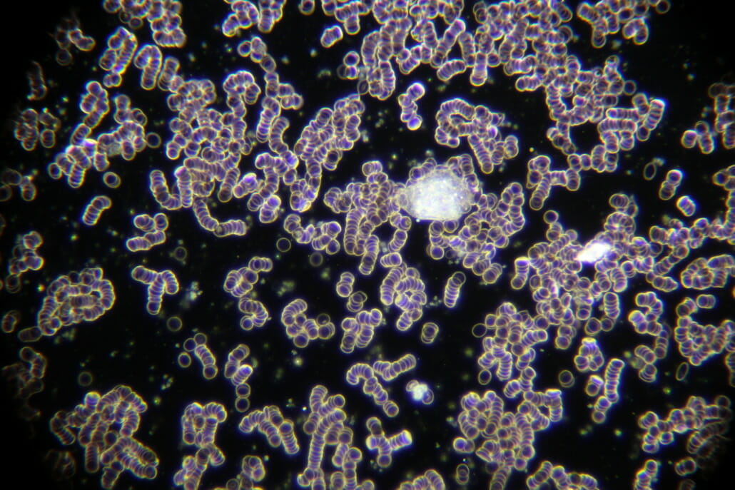
Fig. 1: Images taken under the dark field microscope. You can clearly see the money roll effect of the red blood cells, as caused for example by strong electrosmog exposure.
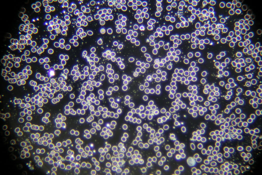
Fig. 2: Images taken under the dark field microscope. In this image, the red blood cells are not jammed and can move freely in an optimal way.
The consequences are:
- poor nutrition of the cells
- clumping of red blood cells
- overacidification of the body.
This is the basis of many diseases such as:
- lack of drive and states of exhaustion
- rheumatism
- thrombosis
- embolism
- infarct.
Dark field microscopy as a method of detecting the Rouleaux effect
With a dark field microscope, these clumps of red blood cells (erythrocytes), known as the money roll effect, can be optically detected after only a few minutes of mobile phone calls.
If the clumps have formed in the blood, the state of exhaustion can subside after hours by staying in a stress-free environment. The blood cell formations get back to normal again. In a light to moderately stressed environment, regeneration may only be insufficient or might not be possible at all.
Waveguard – your certified protection against electrosmog
Take your EMF protection into your own hands! To help you with this, we develop exclusive products to serve you both for on the go and for at home or in the office. Our Qi technology is built into all of our Qi devices.
Benefits of Qi technology:
- use modern technology such as wifi, cellular network & Bluetooth without hesitation;
- German technology & production;
- non electric;
- simple application;
- free regeneration;
- every year, around 2,500 customers worldwide trust Waveguard with their EMF protection.
Do you have any questions about your personal EMF protection? We are here to help.
Did you like this article? Follow the latest news on the subject of electrosmog in our Waveguard blog.
Sources
- Pascoe pharmaceutical preparations GmbH: Dark field microscopy – a holistic blood test. Giessen 2021 https://www.naturheilkunde.de/naturheilverfahren/dunkelfeldmikroskopie.html
- Fachverband Deutscher Heilpraktiker e.V. Bundesverband: Dunkelfeld-Diagnose. Bonn 2021. https://www.heilpraktiker.org/dunkelfeld-diagnose
- International Society for Electrosmog Research IGEF Ltd .: Ruleaux effect in the blood due to electrosmog. Dublin 2017. https://www.elektrosmog.com/gesundheit/elektrosmog-veraendert-ihr-blut
- Klinik im LEBEN GmbH: dark field diagnostics – a holistic blood test. Greiz 2021. https://www.klinik-imleben.de/de/dunkelfelddiagnostik/
- Health Foundation; Doctor’s information: dark field blood test Hamburg 2020. https://www.arzt-auskunft.de/gesundheit/dunkelfeld-blutuntersuchung/
- Naturheilpraxis Katrin Mögel: Vital blood test in the dark field. Dorfhain 2021. https://naturheilpraxis-moegel.de/vitalblutuntersuchung-dunkelfeld/
- Naturheilpraxis Britta Varnhorn: Treatment. Leipzig 2021. https://brittavarnhorn.de/Behandlung/
- International Society for Electrosmog Research IGEF Ltd: Rolls of money in the blood due to electrosmog. Dublin 2017. https://www.elektrosmog.com/gesundheit/elektrosmog-veraendert-ihr-blut
- WELT; Axel Springer SE: Comprehensive diagnosis using dark field microscopy . Berlin 2012. https://www.welt.de/gesundheit/medizin-ratgeber/article157721762/Umfassende-Diagnose-durch-Dunkelfeldmikroskopie.html